
Figure 3. Negative staining of Bac-VP1 associated with bilosomes.
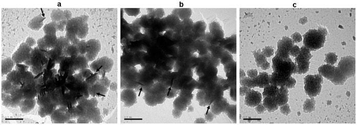
(a) Bac-VP1 associated with bilosomes, (b) Inactive Bac-VP1 associated with bilosomes (c) Bilosomes only. Imaging was carried out using a transmission electron microscope JEM-1230 (Jeol). Arrows pointing to the successful association of Bac-VP1 with bilosomes.