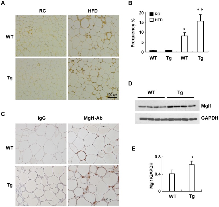
Figure 6. Effect of HFD on macrophage infiltration in visceral adipose tissues of WT and Tg mice.
WT and Tg mice were fed with a RC or HFD for 12 weeks, and then sacrificed. A, The WAT sections were subjected to immunostaining using antibody against F4/80 as described in Materials and Methods. B, F4/80-positive macrophages were quantified as the percentage of adipocytes surrounded by a crown-like structure in the total adipocytes. The number of animals in each group was five to six. *P<0.05 vs the RC-fed group of the same genotype; † P<0.05 vs HFD-fed WT group. C, The WAT sections of HFD-fed WT and Tg mice were immunostained with antibody against Mgl1 as described in Materials and Methods. D, The Mgl1 expression levels in WAT tissues of HFD-fed WT and Tg mice were examined by Western blot analysis. E, The Mgl1 protein levels were quantified by densitometry. The number of animals in each group was four. *P<0.05 vs the WT group.

