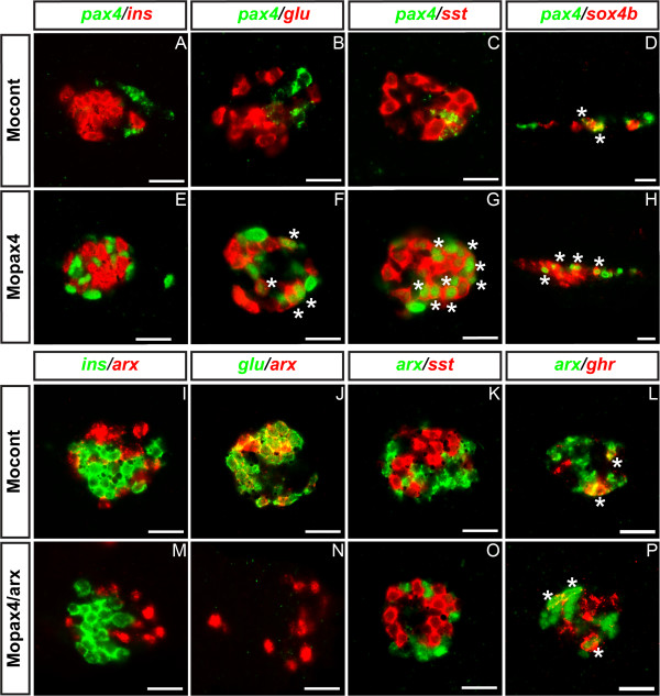
Figure 8.
Identity of pax4+ and arx+ cells following Pax4 and Arx inactivation. (A – F) Double fluorescent WISH performed on 30 hpf embryos showing expression of pax4 transcripts (green) in the different pancreatic cell types of control morphants (A, C, E, G) and in pax4 morphants (B, D, F, H). All panels display ventral vues except panels D and H which are lateral vues. (I – N) Confocal images of pancreatic area with arx probe and various pancreatic hormones at 30 hpf in control (I-L) and double pax4/arx morphants (M-P). Anterior part to the left. Scale bar = 20 μm. Asterisks (*) indicate double positive cells.

