
Figure 2. Subcellular distribution of human PDZ domains when fused to eYFP-S1PDZ1.
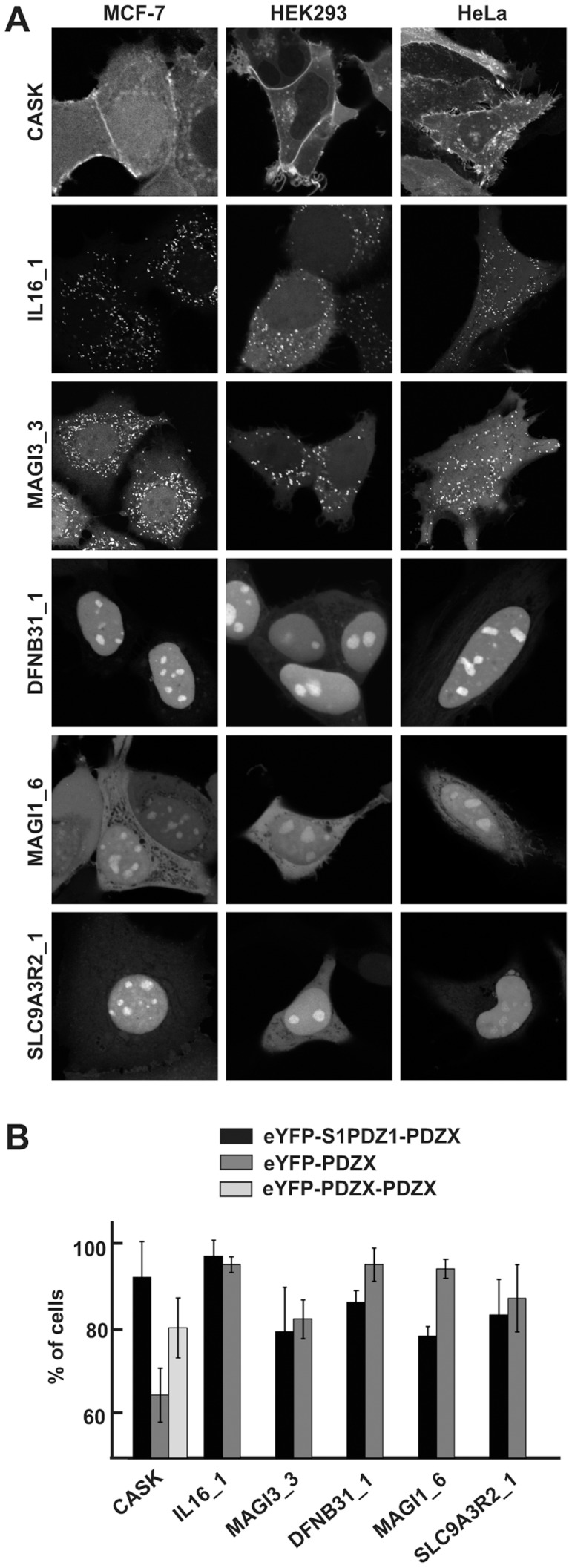
A. Confocal micrographs of MCF-7, HEK293 and HeLa cells transiently over-expressing selected eYFP-S1PDZ1 tagged PDZ domains, as indicated. Shown are examples of strong plasma membrane localization (CASK), bright cytosolic spots (MAGI3_3 and IL16_1) and enrichment in subnuclear organelles (DFNB31_1, MAGI1_6 and SLC9A3R2_1). B. Bar graph illustrating the mean percentage of MCF-7 cells (± S.D.) where the eYFP-S1PDZ1-tagged and eYFP-tagged PDZ domains were enriched in the characteristic subcellular compartments.