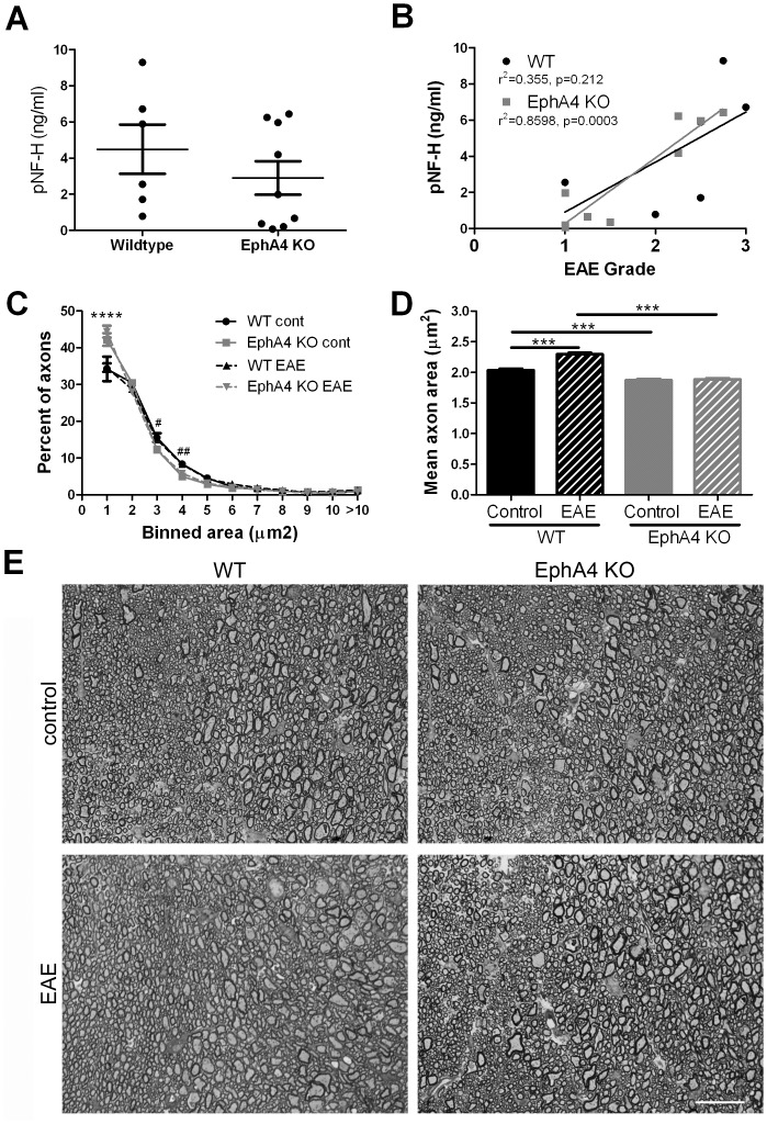
Figure 6. Axonal damage in EAE-affected wild-type and EphA4 knockout spinal cords.
(A,B) Blood samples from EAE-affected wild-type (WT, n = 6) and EphA4 knockout (KO, n = 9) mice were taken at 20 days post-immunisation and pNF-H levels were measured as an indicator of axonal injury. A) There was no significant difference in mean pNF-H levels between genotypes. B) In EphA4 knockout mice there was a significant correlation between pNF-H levels and highest clinical score reached per mouse; a similar non-significant trend was observed in wild-type mice. C,D) Analysis of axonal diameter in the dorsal funiculus of control and EAE-affected mice indicated that KO mice had a greater number of small diameter axons than wildtype control and EAE-affected WT mice. C) The distribution of axon diameters did not differ between control and EAE-affected mice but WT mice had fewer axons in the smallest category in either case. 2-way ANOVA p<0.0001, ****p<0.0001 and ##p<0.01, #p<0.05 for EAE-affected mice only. D) The mean diameter of WT but not KO axons increased with EAE compared to control mice, indicative of increased axonal pathology in the wildtype mice and a relative sparing of KO axons. 1-way ANOVA p<0.0001, ***p<0.001. E) Representative images of axons in the dorsal funiculus in normal control and EAE-affected WT and EphA4 KO mice. Scale bar 20 µm.

