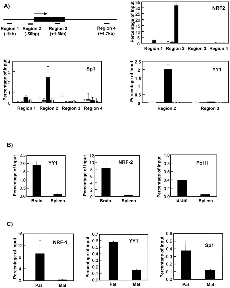
Figure 7. ChIP analysis of the Mkrn3 and Ndn promoters.
A) ChIP analysis of the Mkrn3 locus. Antibodies against NRF-2, Sp1, and YY1 were used to immunoprecipitate chromatin from the maternal and paternal alleles separately in TgPWSdel and TgASdel mouse fibroblasts, respectively. The location of primers used to examine transcription factor binding within regions 1–4 across the Mkrn3 locus are described further in the main text. The solid rectangle depicts the intronless Mkrn3 gene; the bent arrow represents the transcription initiation site. Open bars represent analysis of the maternal allele, bars with horizontal stripes represent control samples from TgPWSdel cells (maternal allele) treated with no antibody, solid bars represent analysis of the paternal allele, and bars with vertical stripes represent control samples from TgASdel cells (paternal allele) treated with no antibody. B) ChIP analysis of the Mkrn3 promoter region (region 2 in panel A) in primary mouse brain and spleen cells. Brain and spleen cell preparations from C57BL/6 mice were subjected to ChIP analysis with antibodies against YY1, NRF-2, or RNA polymerase II. C) ChIP analysis of the Ndn promoter region in TgPWSdel and TgASdel cells using antibodies against NRF-1, YY1, and Sp1.

