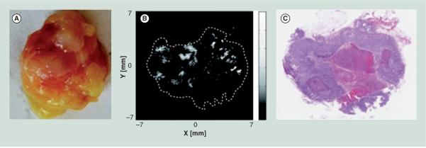
Figure 2. Images of a resected human lymph node showing photoacoustic signal corresponding to locations within the tumor that contain melanoma cells.

(A) Photograph of the resected lymph node. (B) Photoacoustic signal acquired from the approximate center of the node. (C) Hematoxylin and eosin stained histopathological section of the approximate center of the node, with the dark regions indicating melanoma cells and the light region in the center indicating an area of necrosis.
Reproduced with permission from [74] © Society of Photo Optical Instrumentation Engineers (2011).
