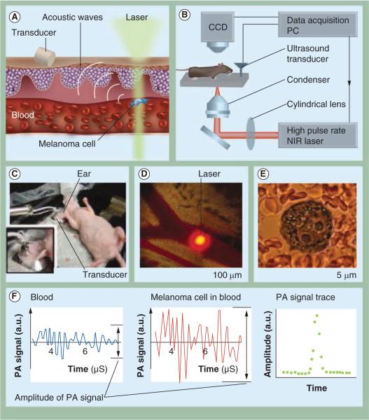
Figure 3. Description of an in vivo photoacoustic flow cytometer.
The principle of operation for the detection of melanoma circulating tumor cells (A), the instrumentation set up (B), the in vivo experimental set up (C) and image of a mouse ear blood vessel and the laser beam (D), an image of a melanoma cell within the vessel (E) and the resulting photoacoustic signals (F) from blood (left) from a single B16F10 melanoma cell within blood (middle), and the photoacoustic signal trace in the presence of a melanoma cell (right) (F) are all shown.
CCD: Charge-coupled device; NIR: Near-Infrared; PA: Photoacoustic; PC: Personal computer.
(A)&(B): Modified with permission from [87]. (C)–(F): Reproduced with permission from [87] © International Society for Advancement of Cytometry (2011).

