Figure 2.
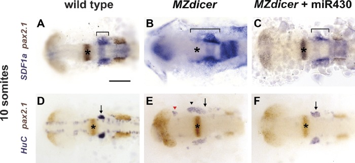
miR-430 refines SDF1a mRNA expression. 10-somite-stage embryos were stained for SDF1a (blue, A–C) or HuC (blue, D–F) and pax2.1 (brown) mRNA to visualize the SDF1a mRNA expression domain (A–C) or TgSNs (D–F) in relation to the MHB (asterisks), respectively. The bracket marks the anterior–posterior extent of SDF1a mRNA expression in A–C. Arrows and arrowheads denote correctly positioned and mispositioned TgSNs, respectively, in D–F. The red arrowhead in E denotes mispositioned neurons located close to the eye. Dorsal view, anterior to the left. (A and D) Uninjected wild-type embryos. (B and E) Uninjected MZdicer mutant embryos. (C and F) MZdicer mutant embryos injected with miR-430 RNA. Bar, 100 µm.

