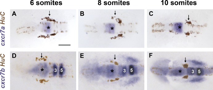
Figure 4.
cxcr7a and cxcr7b are expressed during TgSN migration. (A–C) cxcr7a (blue) and HuC (brown) mRNA distribution in 6-, 8-, and 10-somite-stage embryos. HuC stains TgSNs (arrows). (D–F) cxcr7b (blue) and HuC (brown) mRNA distribution in 6-, 8-, and 10-somite-stage embryos. Dorsal view, anterior to the left. MHB is labeled with asterisks. Hindbrain rhombomeres 3 and 5 are indicated by “3” and “5.” Bar, 100 µm. See also Fig. S2.

