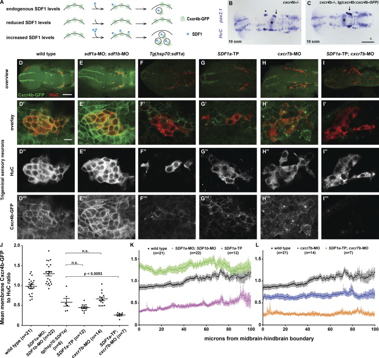
Figure 9.
TgSNs are exposed to elevated SDF1 protein levels when Cxcr7b or miR-430 activity is reduced. (A) Conceptual overview of quantification of SDF1-Cxcr4b signaling. (B) 12-somite cxcr4b−/− nontransgenic sibling embryo stained in blue for HuC and pax2.1 mRNA to visualize TgSNs in relation to the MHB (asterisks). (C) 12-somite cxcr4b −/−; tg(cxcr4b:cxcr4b-GFP) sibling embryo. Arrows and arrowhead denote correctly and incorrectly positioned TgSNs, respectively. (D–I) Overview of a representative embryo of the indicated genotype stained for TgSNs with HuC (red) and for Cxcr4b-GFP (green). (D′–I‴) High magnification of embryos stained as indicated in D–I. (D′–I′) Overlay of Cxcr4b-GFP and HuC Fluorescence. (D″–I″) HuC fluorescence. (D‴–I‴) Cxcr4b-GFP fluorescence. All embryos are eight somites. (D–D‴) Wild-type embryo. (E–E‴) SDF1a and SDF1b morpholino–injected embryo. (F–F‴) Heat-shocked tg(hsp70:SDF1a) embryo. (G–G‴) SDF1a-TP embryo. (H–H‴) cxcr7b morpholino–injected embryo. (I–I‴) SDF1a-TP and cxcr7b morpholino–injected embryo. (J) Mean Cxcr4b-GFP/HuC ratios ± SEM on the outer membranes of TgSNs in indicated genetic scenarios at the eight-somite stage. All differences between the different genetic scenarios are statistically significant, with P < 0.0001, except where noted otherwise. n.s., not significant. (K–L) Mean Cxcr4b-GFP/HuC ratios ± SEM on the outer membranes of TgSNs along the migration route in the indicated genetic scenarios at the eight-somite stage. Because of the neuron dispersal in heat-shocked tg(hsp70:SDF1a) embryos, quantification could not be resolved along the migratory route. 0 µm corresponds to the MHB. Bars: (B and C) 100 µm; (D–I) 100 µm; (D′–I‴) 10 µm.

