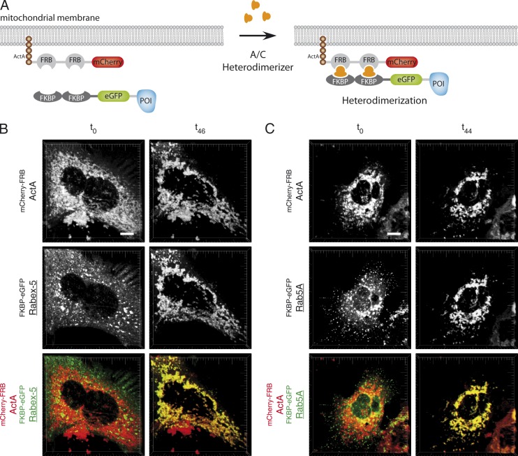
Figure 2.
Mistargeting of Rab5A and Rabex-5 to mitochondrial membranes. (A) Domain structure of fluorescent fusion proteins. The scheme depicts the mistargeting of the protein of interest (POI) through A/C heterodimerizer–induced dimerization of FRB and FKBP domains. (B and C) Confocal images of live cells showing the A/C heterodimerizer–induced translocation of FKBP-eGFPRabex-5 (B) and FKBP-eGFPRab5A (C) to mitochondria in Cos-7 cells cotransfected with mitochondrially anchored mCherry-FRBActA. Addition of A/C heterodimerizer to a final concentration of 1 µM, 24 h after transfection, leads to the targeting of Rabex-5 (B) or Rab5A (C) from the cytosol and endosomes to mitochondria as seen by the appearance of yellow areas in the merged image indicating colocalization. Bars, 10 µm.

