
Figure 2. Expression of DPPX in myenteric plexus.
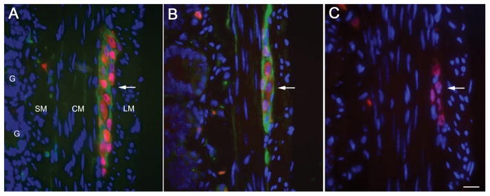
Transverse section of small bowel of rat showing the longitudinal muscular layer (LM), circular muscular layer (CM), submucosal layer (SM), and glans (G). The myenteric plexus (Plex) is revealed as clusters of large neurons between the two muscular layers. In the 3 panels (A–C) the nuclei of the neurons (red) was labeled with anti-Hu (a highly specific neuronal marker). Panel A, shows in green the DPPX immunostaining using a rabbit polyclonal antibody (1:1000, developed by BD); panel B shows the DPPX reactivity of serum from one of the patients with encephalitis, and panel C shows the lack of reactivity of serum from a healthy subject. Note that DPPX is predominantly expressed in the cytoplasm-membrane of the large clustered neurons of the myenteric plexus, and is also detected in a fine longitudinal pattern in CM and SM where the submucosal plexus is located. Bar = 20μm.