Figure 2.
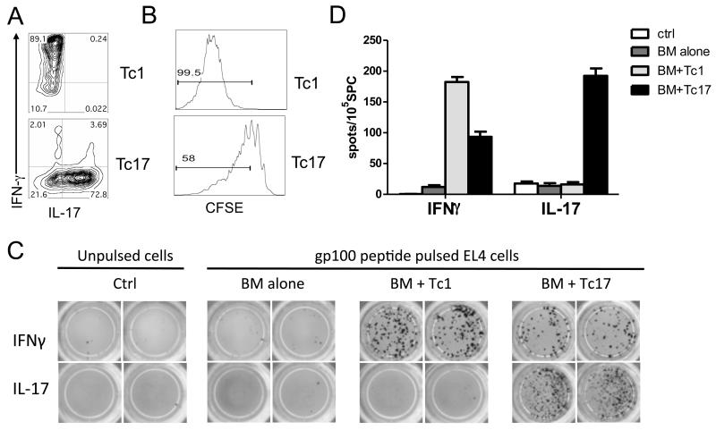
Generation and Tc1 and Tc17 Responses of antigen-specific to tumor antigen stimulation ex vivo. (A) Tc1 and Tc17 cells were generated as described in “Materials and Methods”, and their phenotype in terms of IFNγ and IL-17 production was shown on gated Pmel-1 (Thy1.1+) cells. (B) In a separate experiment, pmel-1 Tc1 and Tc17 cells were labeled with CSFE and transferred into B16 tumor-bearing B6 mice (n = 4). Five days after cell transfer, mice were euthanized and their splenocytes were stained for CD8 and Thy1.1. The CSFE profile is shown on gated live CD8+Thy1.1+ cells for one of four representative mice. (C) A set of B6 mice (n = 3) were inoculated i.v. with luciferase transduced B16 F10 cells and received Tc1 or Tc17 cells combined with BMT. Mice were euthanized on day 14 after treatment. Splenocytes were isolated and processed for cytokine-release ELISPOT assays. Gp100-specific IFNγ- and IL-17-producing cells were detected after co-culture with gp100-pulsed and unpulsed EL4 for 20 hours. Experiments were repeated 2 times with similar results. (D) The graph shows the average number of IFNγ- and IL-17-producing spots from triplicate wells with 1SD as error bars.

