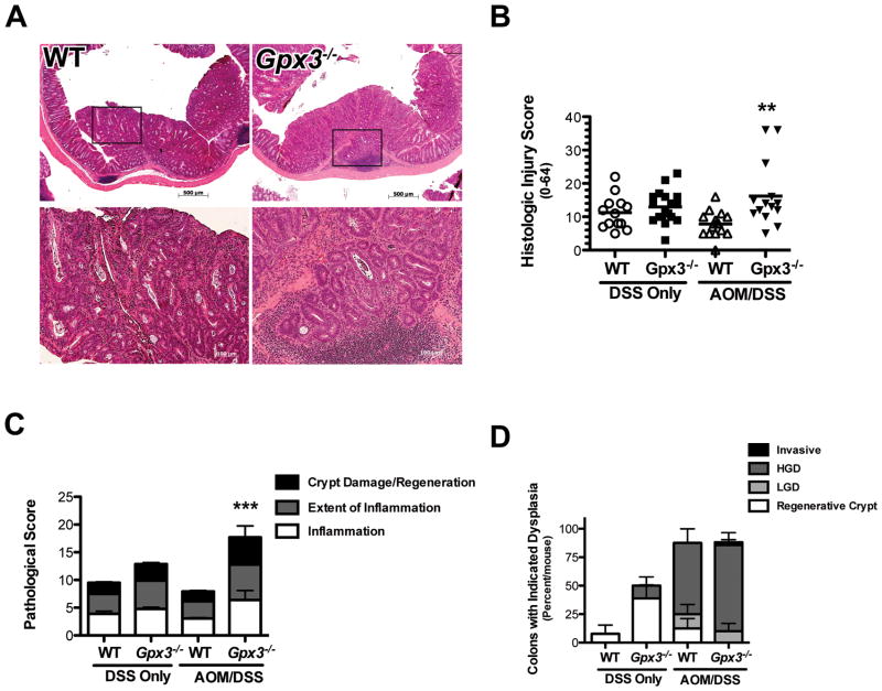
Figure 3. Increased histologic injury in Gpx3−/− colons.
A) Representative H&E staining from WT (10x top, 20x bottom left) or Gpx3−/− (10x top, 20x bottom right) colons. There is evidence for invasive adenocarcinoma in the Gpx3−/− tumor. B) Histologic injury score for DSS only or AOM/DSS tissues. ** P<0.01. C) Division of the histological injury scores from Fig. 3B into pathological inflammation, extent, and crypt damage/regeneration scores for DSS only or AOM/DSS tissues. ***P<0.001. D) Dysplasia grading (performed by MKW) in WT and Gpx3−/− tumors. Results represent percentage of total tumors for each group within each grade.

