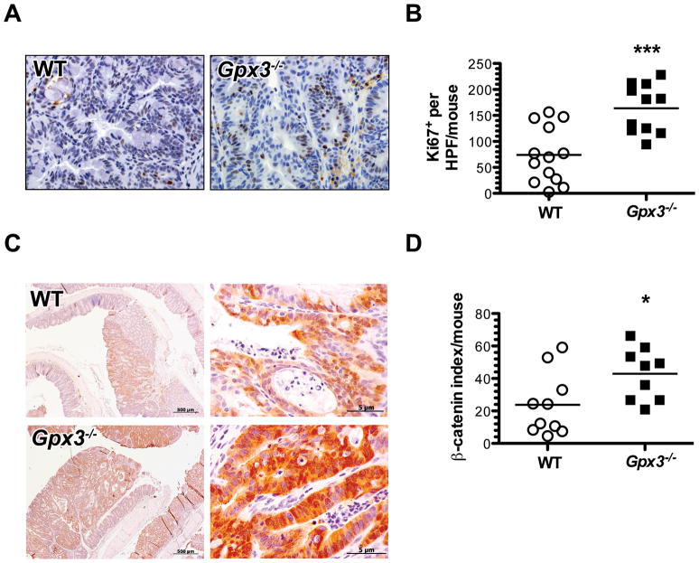
Figure 4. Increased intratumoral proliferation and nuclear β-Catenin in Gpx3−/− tumors.
α-Ki67 immunohistochemistry was performed to identify actively proliferating cells. A) Representative images of Ki67 staining in WT or Gpx3−/− tumors (40x magnification). B) Intratumoral proliferation index calculated from number of BrdU positive cells per HPF in 20 HPF/mouse. ***P=0.0003. C) β-catenin expression and localization was determined via immunohistochemistry with β-catenin as per Methods section (Gpx3−/−, N=10, WT, N=10 tumors). Representative staining for β-catenin from WT or Gpx3−/− tumors (left, 10x magnification; right, 40x magnification). D) Intratumoral β-catenin index calculated as described in the Methods section. *P=0.03.

