Figure 1.

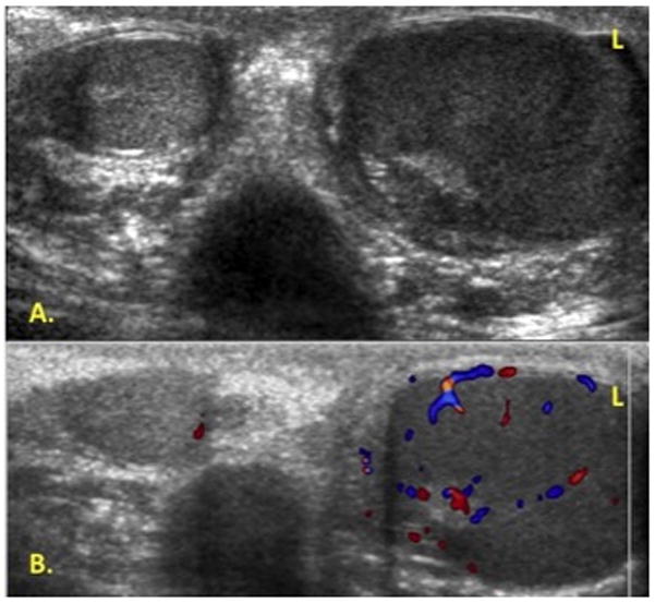
Scrotal doppler ultrasound. A. Asymmetrically enlarged hypoechoic left testicle compared to right. B. Left testicle with increased vascularity without focal mass.

Scrotal doppler ultrasound. A. Asymmetrically enlarged hypoechoic left testicle compared to right. B. Left testicle with increased vascularity without focal mass.