Figure 2.

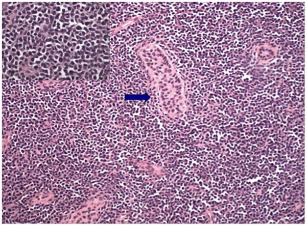
Testicular biopsy demonstrating extensive and diffuse infiltrate of lymphoblasts, residual tubule, arrowed, (Haematoxylin & Eosin stain, ×100). Higher magnification, insert, H&E stain, x400

Testicular biopsy demonstrating extensive and diffuse infiltrate of lymphoblasts, residual tubule, arrowed, (Haematoxylin & Eosin stain, ×100). Higher magnification, insert, H&E stain, x400