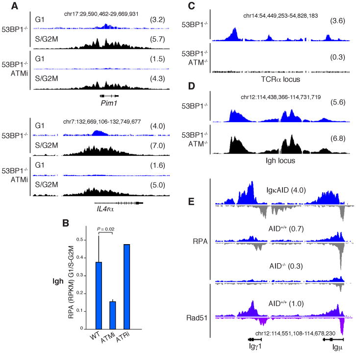
Figure 5. ATM is required for G1 but not S-G2/M resection.
(A) RPA accumulation at Pim1 and IL4Rα loci from 53BP1−/− activated B cells that were either treated (lower two panels) or not treated (upper panels) with the ATM inhibitor KU-55933. Samples were sorted into G1 or S/G2/M phased cells using the Hoechst dye 33342. Numbers in parenthesis represent RPKM values within the specified genomic windows. (B) RPA accumulation at Igμ and Igγ1 loci in WT, ATMi-, or ATRi-treated B cells. Values represent the RPKM ratio between G1 and S-G2/M-phased cells. (C–D) RPA recruitment to the TCRα in thymocytes (C) or Igh in activated B cells (D) from 53BP1−/− (upper) or 53BP1−/−ATM−/− (lower) mice. (E) RPA and Rad51 recruitment to activated B cells with an intact NHEJ: IgκAID transgenics, AID+/+, and AID−/−.

