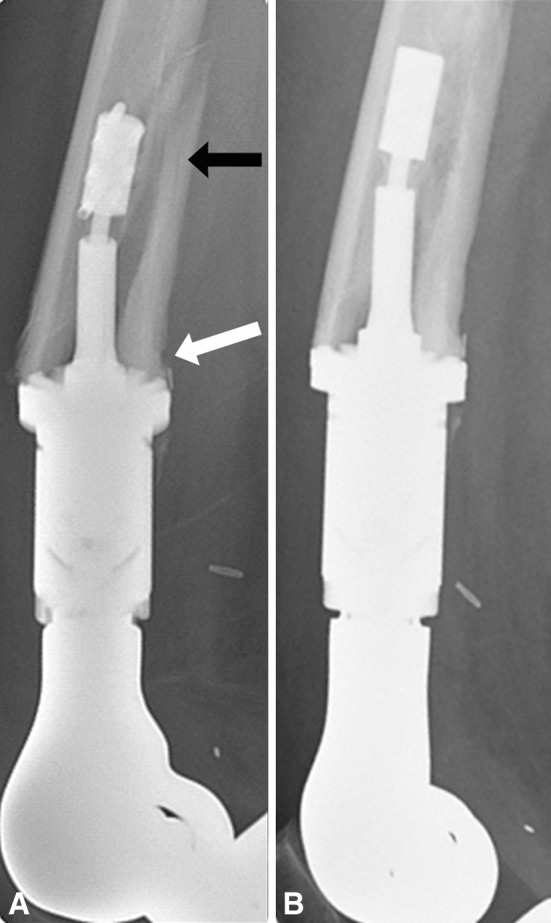
Fig. 7A–B.

(A, B) Lateral radiographs taken 3 months apart show a Type IIB failure. (A) The black arrow points to the fracture of the posterior cortical segment that has moved with the spindle and the megaprosthesis, and the white arrow points to the intact bone-spindle interface where there has been some bone hypertrophy. Notably, the anterior cortex has not integrated and there is no hypertrophy. (B) The fracture healed spontaneously.
