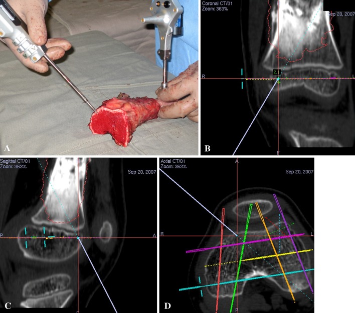
Fig. 7A–D.
(A) A photograph shows the tumor specimen for Patient 4 after computer-assisted joint-preserving resection. The image-to-patient registration remains valid as the dynamic reference tracker is still attached to the tumor specimen after resection. The tip of the navigation probe is on the distal bone end of the tumor specimen. The corresponding (B) coronal, (C) sagittal, and (D) axial views on the navigation monitor are shown. The virtual tip of the navigation probe (red cross) is exactly at the resection level virtually planned on the preoperative CT images. The distance between the achieved resection (at the virtual tip of the navigation probe) and planned resection (at the virtual pedicle screws) is measured. For a better illustration, see Video 2 (supplemental materials are available with the online version of CORR®).

