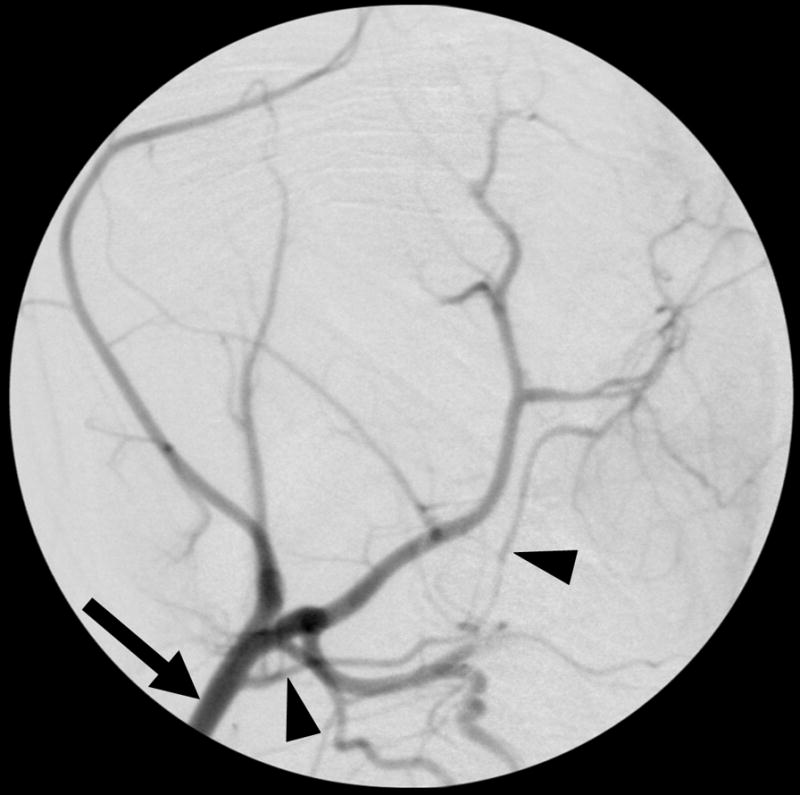
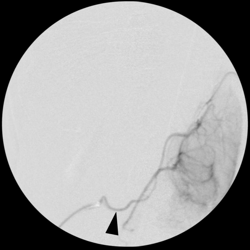
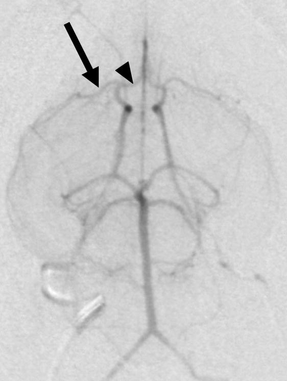
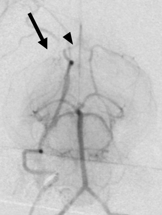
Figure 1. Selective rabbit intracranial angiography.


(a) Common carotid selection (arrow), lateral view. The internal carotid artery is seen coursing to the base of the brain (arrowheads). (b) Internal carotid sub-selection (arrowhead) in a lateral view demonstrates filling of the cerebral vasculature. (c) A frontal-view internal carotid angiogram clearly shows the Circle of Willis including the middle cerebral artery (MCA, arrow), and the anterior cerebral artery (ACA, arrowhead). (d) Repeat angiography after injection of three embolic microspheres (700–900 μm) shows the occlusion of the MCA (arrow) and persistent flow in the ACA (arrowhead).
