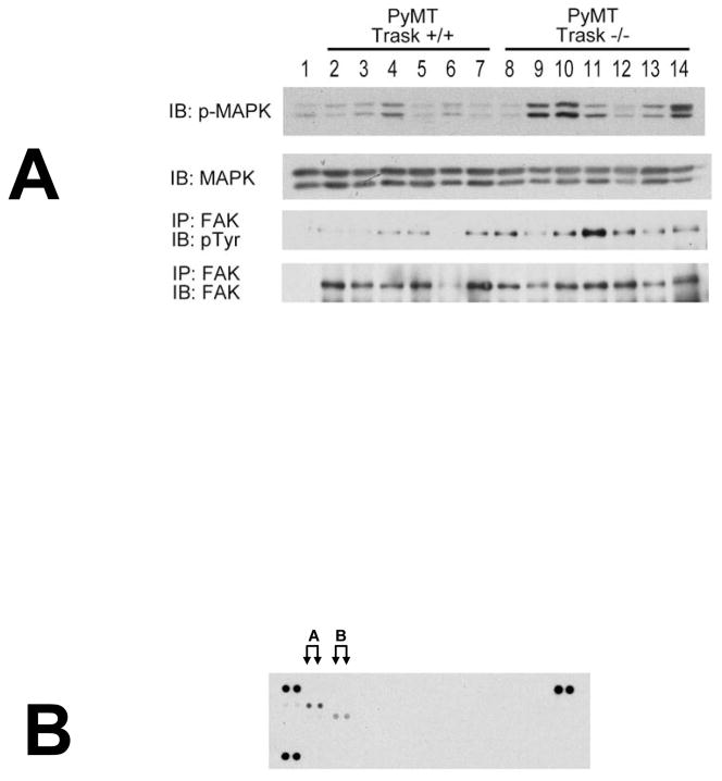
Figure 4. Integrin and growth factor signaling in PyMT tumors in vivo.
(A) Lysates from PyMT Trask +/+ and PyMT Trask −/− tumors were analyzed by immunoblotting. Phosphorylation of MAPK (T202/Y204), tyrosine phosphorylation of FAK and their total protein expressions are shown. Each lane corresponds to a tumor derived from a different mouse. (B) The Trask +/+ tumor lysates were used in the R&D Systems p-RTK profiling. This analysis revealed that the predominant active RTK in these tumors is HER2 (labeled A) and to a lesser extent PDGFR (labeled B).

