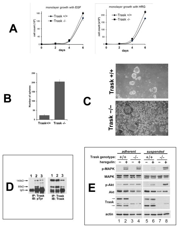
Figure 5. Loss of Trask augments growth factor signaling in PyMT tumor derived cell lines.
(A) Tumor cell lines from MMTV-PyMT mice in the indicated Trask genotypes were grown in the presence of either epidermal growth factor or heregulin for six days and counted. (B) Tumor cell lines from PyMT-driven mouse mammary tumors originating from Trask+/+ or Trask−/− mice were cultured in non-adherent plates for 7 days and the number of spheroids counted manually under microscopic examination. (C) Phase contrast microscope images of the spheroids are shown here. (D) Cell lysates were assayed for the expression and phosphorylation of Trask by immunoprecipitation. Lanes correspond to 1) PyMT tumor cell line cultured in the adherent state, 2) PyMT tumor cell line cultured in the suspended state, 3) in vivo PyMT tumor from a mouse. Trask is phosphorylated during tumor growth in vivo, resembling the suspended state when grown in culture. (E) Serum starved PyMT cell lines from the indicated genotypes were grown in adherent or suspended form were stimulated with heregulin. The growth factor-induced activation of MAPK is suppressed in the suspended Trask+/+ cells. This suppression does not occur in Trask−/− cells. There is a more subtle enhancement of Akt signaling in the Trask−/− cells. The arrows indicate the 140 and 80kD forms of Trask. The phospho-immunoblots reflect the T202/Y204 phosphorylation of MAPK and the S473 phosphorylation of Akt.

