
Fig. 2.
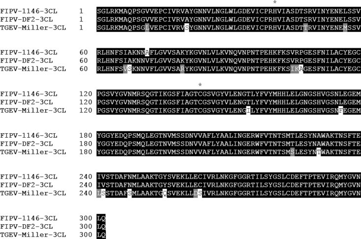
The sequence alignment of the 3CL protease of TGEV (Miller strain) and feline coronaviruses (WSU-1146 and DF2 strains). Asterisks mark the position of the conserved cysteine (144) and histidine (41) residues in the active site.
Official websites use .gov
A
.gov website belongs to an official
government organization in the United States.
Secure .gov websites use HTTPS
A lock (
) or https:// means you've safely
connected to the .gov website. Share sensitive
information only on official, secure websites.
The sequence alignment of the 3CL protease of TGEV (Miller strain) and feline coronaviruses (WSU-1146 and DF2 strains). Asterisks mark the position of the conserved cysteine (144) and histidine (41) residues in the active site.