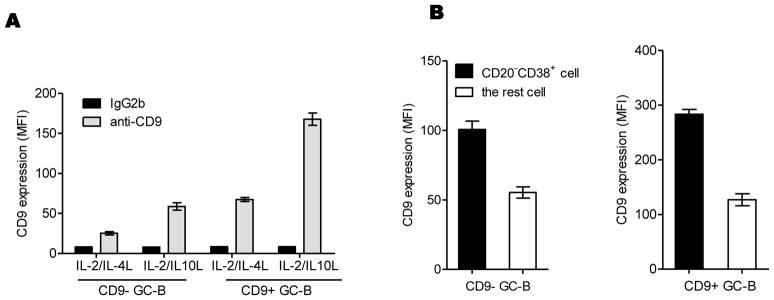
Figure 3. CD9 is induced during GC-B cell differentiation to PC.

CD9− and CD9+ GC-B cells were cultured in the presence of FDC/HK cells with a different combination of cytokines (CD40L+ IL-2 + IL-4 or CD40L+ IL-2 + IL-10) for 4 days. (A) At the end of the culture, cells were stained with anti-CD9 or isotype control antibody. CD9 expression levels on the cells were quantified by FACS analysis and shown as MFIs. (B) Cells generated from the culture with CD40L+ IL-2 + IL-10 were further stained with anti-CD20 and anti-CD38, gated by the expression of these two markers, and the expression of CD9 on CD20-CD38+ plasmablasts and the rest was also quantified by FACS analysis and presented in MFIs. One representative data from three independent experiments with different donors is shown.
