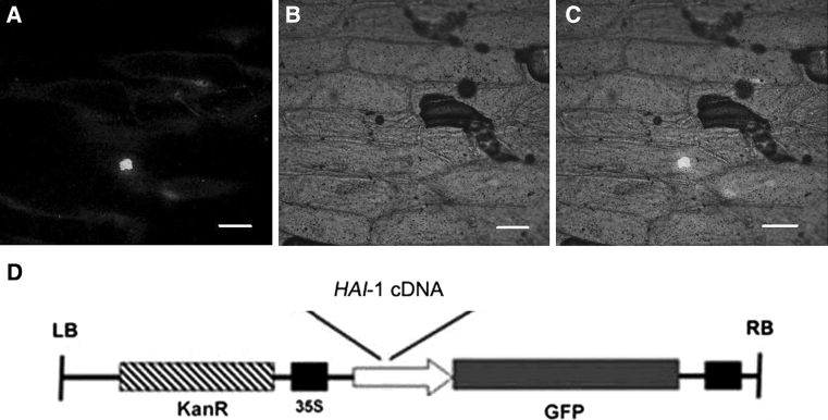
Fig. 10.
Transient expression of HAI-1-GFP in the bombarded onion epidermal peels. The HAI-1-GFP fluorescence was found to be localized to the nucleus. a–c Fluorescence image, bright field image and merged fluorescence image of 35S::HAI-1-GFP. Bars 100 μm. d The schematic diagram of HAI-1:GFP fusion vector

