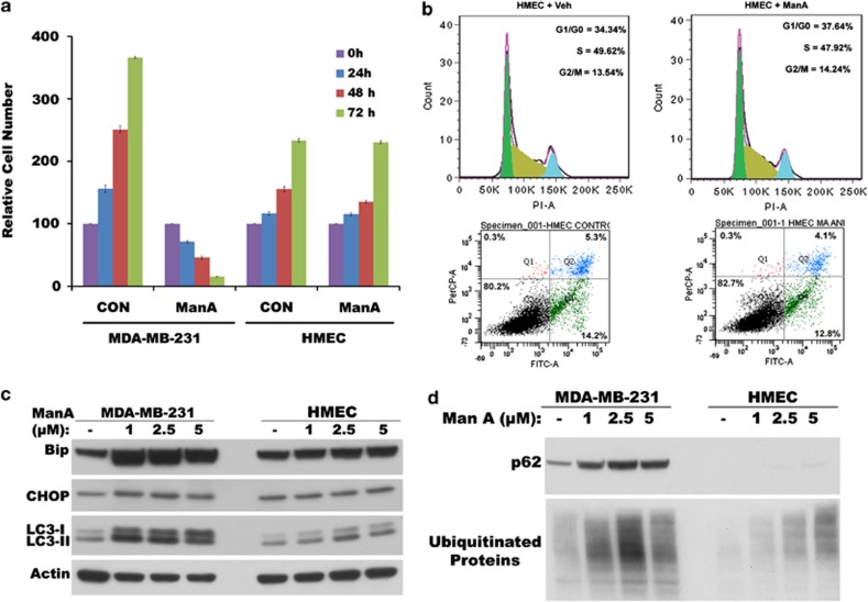
Figure 4.
Normal HMEC are protected from Man A-induced cytotoxic effects. (a) Time course of cell viability measured by MTT assay in MDA-MB-231 cells and HMEC after treatment with 5 μM Man A. (b) Cell cycle analysis by FACS after propidium iodide staining (top panel) and analysis of surface changes and cell permeability by FACS using Alexa Fluor 488 AnnexinV/ dead cell apoptosis kit (bottom panel) in HMEC treated with vehicle (Veh) or Man A for 24 h. (c) Western blots of total cell lysates on the expression of Bip and CHOP, and expression and processing of LC3 in Man A-treated MDA-MB-231 cells and HMEC. (d) Western blots of total cell lysates for p62 expression and accumulation of ubiquitinated proteins in Man A-treated MDA-MB-231 cells and HMEC

