Figure 7.
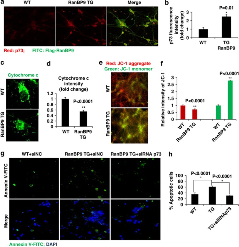
p73 is essential for RanBP9/Aβ1-42-induced apoptosis and mitochondrial dysfunction in primary hippocampal neurons. (a, b) RanBP9 transgenic (TG) and littermate non-transgenic (WT) DIV14 primary hippocampal neurons were subjected to immunostaining using anti-FLAG M2 and anti-p73 antibodies, and images were captured by confocal microscopy. Representative images are shown. Endogenous p73 fluorescence intensity (red) was quantified using Nikon NIS-Elements-AR software (n=4 each). Error bars represent S.E.M. (c,d) RanBP9 transgenic (TG) and littermate non-transgenic (WT) DIV14 primary hippocampal neurons were treated with 0.1% saponin (5 min on ice) to release cytosolic content prior to fixation and subjected to immunostaining using anti-cytochrome c, and images were captured by confocal microscopy. Representative images are shown. (d) Endogenous cytochrome c (FITC) intensities were quantified using Nikon NIS-Elements-AR software (n=4 each). Error bars represent S.E.M. (e,f) Wild-type and RanBP9 transgenic littermate hippocampal primary neurons were subjected to JC-1 staining prior to fixation, and JC-1 signals were captured by fluorescence microscopy. (f) Red (JC-1 aggregate) and green (JC-1 monomer) fluorescence intensities from were quantified using NIS-Elements AR 3.2 software and relative quantifications are shown in the graph (n=4 each). Error bars represent S.E.M. (g,h) Wild-type and RanBP9 transgenic littermate hippocampal primary neurons were transfected with control or p73 siRNA. After 48 h, cells were treated with 1 μM Aβ for 24 h and subjected to staining with Annexin V-FITC and DAPI. Cells were captured directly using fluorescence microscopy. Numbers of Annexin V-FITC-positive cells among DAPI-positive cells were quantified using Nikon NIS-Elements-AR software. The graph shows % Annexin V-positive apoptotic cells (n=3 each). Error bars represent S.E.M.

