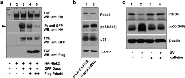
Figure 4.
Pdcd4 inhibits Ser-46 phosphorylation of p53. (a) QT6 cells were transfected with the indicated combinations of expression vectors for HA-Hipk2, GFP-Daxx and Flag-Pdcd4, as indicated below the lanes. Cells were lysed after 24 h and TCEs were either analyzed directly by SDS–PAGE and western blotting with the indicated antibodies or were first immunoprecipitated with antibodies against GFP (second panel from top) before western blot analysis. Hipk2 co-precipitated via Daxx is marked by an arrowhead. (b) MCF7 cells were treated with Pdcd4-specific or control siRNA for 48 h. TCEs were subsequently analyzed with antibodies against Pdcd4, p53 and β-actin. In addition, p53 was first immunoprecipitated from the cell extracts and the immunoprecipitates were then analyzed by western blotting with phospho-p53- (Ser-46) specific antibodies. (c) HEK293 cells were UV irradiated (50 J/cm2) in the presence or absence of caffeine (concentration 6 mℳ). Unirradiated cells served as control. At 4 h after irradiation, TCEs were analyzed by western blotting with antibodies against Pdcd4, phospho-p53(Ser-46) and β-actin.

