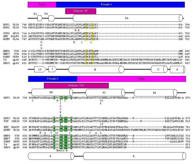
Figure 2.
Protein sequence alignment of herpesvirus and bacteriophage polymerase finger domains. Herpesvirus and bacteriophage polymerase were aligned individually using the Muscle algorithm within Geneious [24,25]. The bacteriophage and herpesvirus sequences were then structurally aligned by RAPIDO [26]. Blocks above sequence highlight structural domains of polymerase. The palm domain is in pink. The fingers domain is in blue. The known conserved regions are shown in magenta blocks above sequence [27,28,29,30,31]. Secondary structural elements of HSV1 UL30 are indicated and are number according to Liu et al. (2006). Secondary structural elements of RB69 gp43 are indicated and are numbered according to Wang et al. (1997). Structural motifs are highlighted in sequence. N helix residues are in yellow. Motif B is in green. Mutations that have been associated with resistance to current anti-herpetic drugs are shown below the corresponding residue [32,33]. Resistance mutations are colored using the following scheme: Red: PyrophosphateR (Resistant), Blue: NucleotideR, Green: PyrophosphateR and NucleotideR. Purple: PyrophosphateHS (Hypersensitive), Brown: NucleotideR but PyrophosphateHS.

