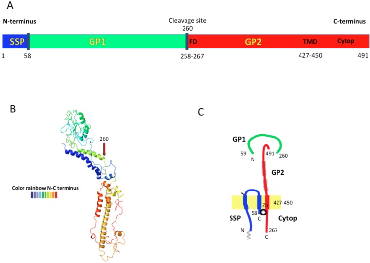
Figure 2.
The LASV envelope glycoprotein precursor (GPC) structure is shown in A) from N- to C-terminus containing SSP, GP1 and GP2 proteins. The dark blue lines represent the cleavage points. The fusion domain (FD); transmembrane domain (TMD), and cytoplasmic domain (Cytop) are shown in brown letters (modified from [19]). B) The predicted Lassa-Josiah GPC structure obtained by open-source software (Phyre2). The structure goes from N-terminus (blue) to C-terminus (red). C) Schematic representation of the trimeric GPC subunit assembled in the cell membrane. GP1 is the most external protein bound to GP2 that is embedded in the lipid membrane. GP2 is thought to interact with SSP through an inter-subunit zinc finger (ball) (modified from [20]).

