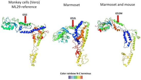
Figure 4.

Predicted ML29 host-specific changes in GPC. The left-most structure shows the ML29 GPC predicted structure after passage in Vero cells. After inoculation into marmosets, the recovered viruses showed an isoleucine (I) to leucine (L) change at position 252 that affects the predicted GPC structure (middle structure). Another mutation at the same position, I to M, also changed GPC structure (figure on the right). The structure goes from N-terminus (blue) to C-terminus (red).
