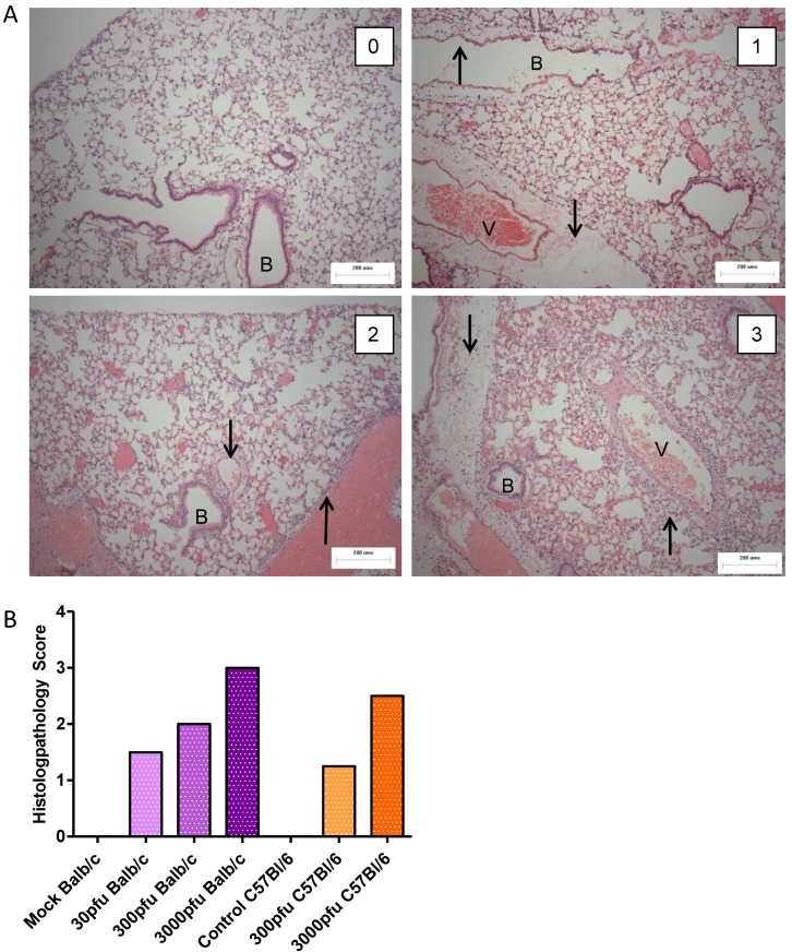
Figure 2.
Histopathological analysis of PVM-infected mice. Five to six week-old Balb/c and C57Bl/6 mice were inoculated with medium, 30 pfu, 300 pfu, or 3000 pfu of PVM 15 and lungs were collected from four mice on day 6 p.i. for histopathological analysis. In (A), representative lung sections for animals scoring 0, 1, 2, and 3 are shown, with the upward arrows (↑) indicating infiltrating inflammatory cells and the downward arrows (↓) indicating oedema in the tissue. The bronchiole is labeled with the letter B and the blood vessel with V. Scores were given on the basis of the severity and dissemination of the lesions visible in duplicate lung sections, and median values are shown for each group (B).

