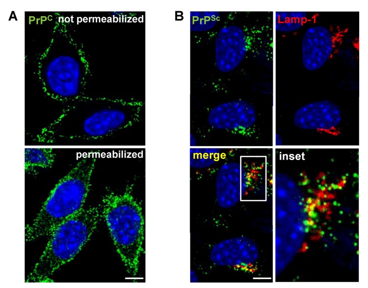
Figure 1.
Localization of PrPC and PrPSc in L929 fibroblast cells. (A) Indirect immunofluorescence (IF) staining of cellular PrP (green) in uninfected L929 cells. PrPC predominantly resides at the cell surface with some intracellular localization. (B) Detection of PrPSc in L929 cells persistently infected with prion strain 22L by IF. In contrast to PrPC, PrPSc (green) primarily localizes intracellularly and partially co-localizes with the lysosomal marker Lamp-1 (red). (A,B) Nuclei were counterstained with Hoechst (blue). Scale bar: 5 µm.

