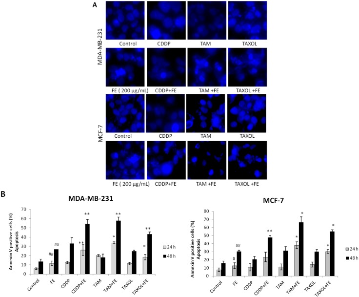
Figure 2.
Synergistic induction of apoptosis by co-treatment. (A) Hoechst 33342 staining of cells treated with 200 μg/mL FE alone or 200 μg/mL FE in combination with 5 μM CDDP, 10 μM TAM or 2.5 nM TAXOL for 48 h. Each experiment shown is representative of 20 randomly observed fields; (B) Analysis of apoptotic cells by annexin/PI double-staining using the IN Cell Analyzer 1000. MDA-MB-231 and MCF-7 cells were treated for different times with 200 μg/mL FE alone or 200 μg/mL FE in combination with 5 μM CDDP, 10 μM TAM or 2.5 nM TAXOL. All results were obtained from three independent experiments. A significant difference from control is indicated by p < 0.05 (#) or p < 0.01 (##); a significant difference from single treatments is indicated by p < 0.05 (*) or p < 0.01 (**).

