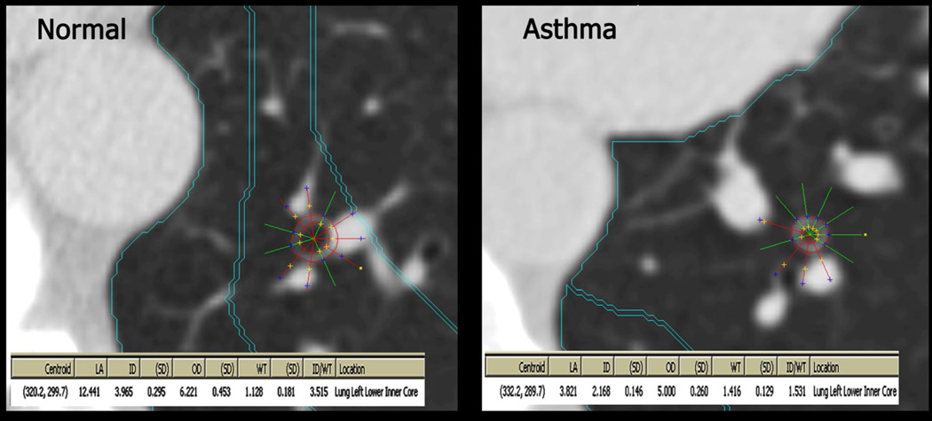
Figure 2. Airway measurements on CT Scan.
Upon locating an airway perpendicular to the plane, a centroid was placed in the airway, from which the Pulmonary Analysis Software Suite generated rays and outer airway diameters. If a ray extended into adjacent tissue the analyst would exclude it and the PASS system would regenerate the inner and outer diameter to conform to the shape of the airway. The remaining rays were averaged to calculate wall thickness and lumen diameter.

