Figure 1.

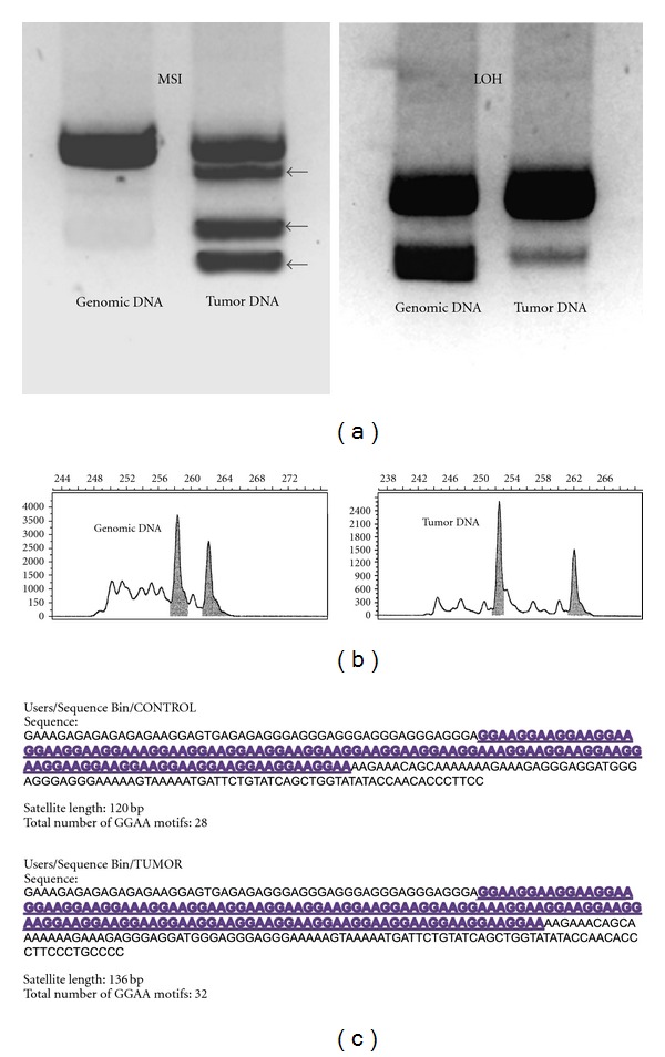
(a) Microsatellite instability was originally assessed using gel electrophoresis and autoradiographic detection. In the left panel, additional bands (black arrows) in the tumor lane illustrate multiple contracted microsatellite alleles relative to the genomic control lane. In the right panel, an information (heterozygous) microsatellite is shown in the genomic control sample, and a significant loss of signal intensity for the smaller allele is observed in the tumor sample, characteristic of allelic imbalance/loss of heterozygosity (LOH). (b) Microsatellite loci are now commonly assessed using fluorescent PCR amplifications, capillary electrophoresis, and automated sequencing techniques. Laser scanners detect fluorescent PCR products and generate a chromatogram displaying microsatellite allele frequencies. Note in the tumor panel, one of the alleles has undergone contraction, depicting MSI in this tumor specimen. (c) Subcloning and direct sequencing of microsatellite amplifications can yield high resolution of the microsatellite sequence and can detect subtle changes in microsatellite constitution. This method of analysis is more time consuming, although programmed bioinformatics software can greatly assist the interpretation of high-volume data. Panel (b) modified with permission from Vilar and Gruber [57].
