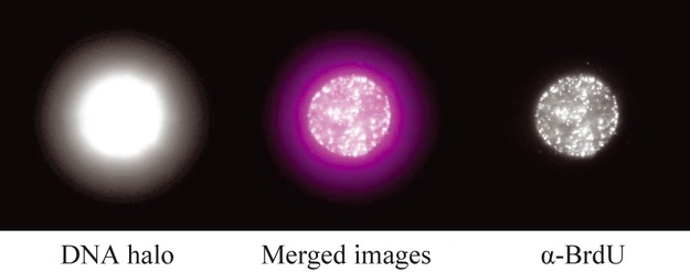
Figure 1.

Newly synthesized DNA (A) Left: example of Maximum Fluorescence Halo Radius image from NIH3T3 cell showing DNA loops stained with DAPI emanating out from the nuclear matrix (NM) (MFHR method described in Buongiorno-Nardelli et al. 1982; Guillou et al. 2010). Right: newly synthesized DNA is observed at the NM but not visible in loop DNA. Cells were pulsed for 30 min with BrdU and visualized with α-BrdU. Centre: merged image showing BrdU (newly synthesized DNA) in white and DNA in magenta.
