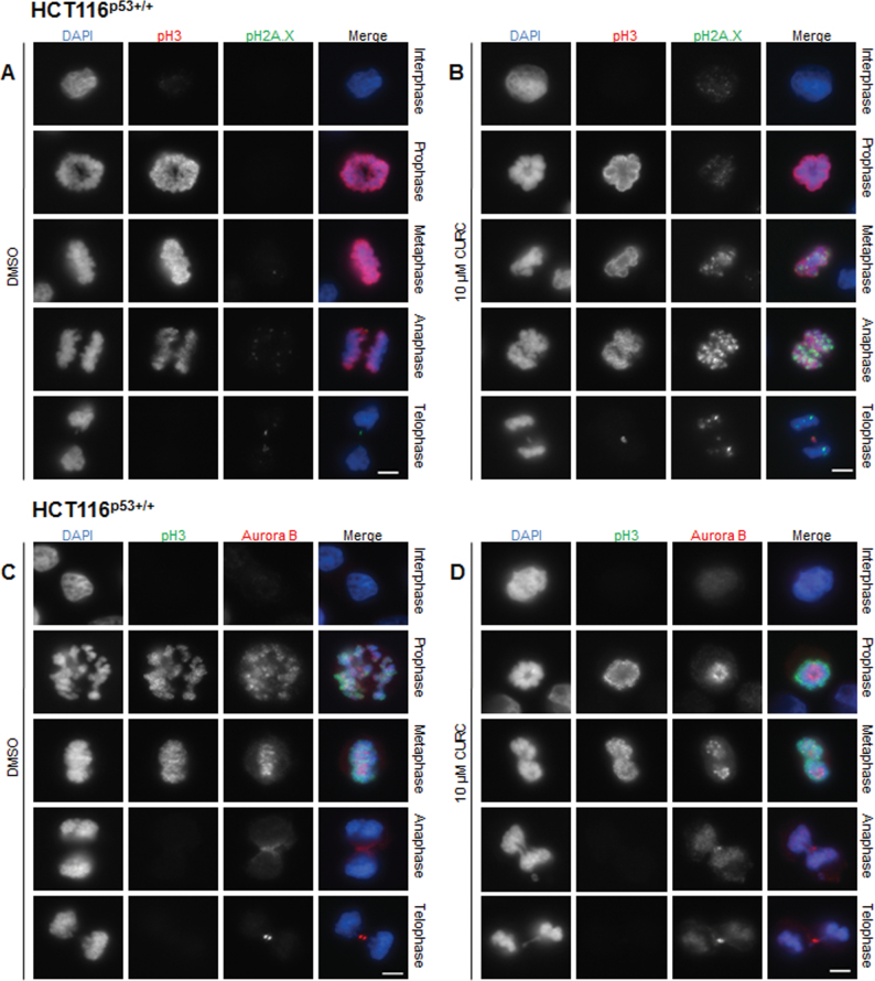
Fig. 6.
Curcumin increased pH2A.X staining and Aurora B mislocalization in mitotic cells. Cells were treated for 12h as indicated and stained with antibodies against pH3 (red) and pH2A.X (green) (A and B) or against pH3 (green) and Aurora B (red) (C and D). DNA was stained with DAPI (blue). Representative images (from n = ≥3 experiments) are shown in interphase prophase, metaphase, anaphase and telophase. Exposure and gain were kept constant for pH2A.X staining. Scale bar, 5 µm.

