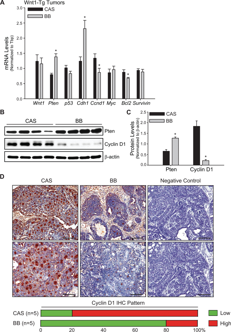
Fig. 3.
Tumors from Wnt1-Tg mice exposed to BB via maternal diet exhibit higher expression of anti-proliferative, proapoptotic and prodifferentiation markers. (A) Genes involved in cell proliferation, apoptosis, differentiation and tumor progression were evaluated by QPCR in tumors from Wnt1-Tg mice exposed to CAS or BB diets in utero and during lactation. Tbp was used as a normalizing control; *P < 0.05 relative to CAS (n = 9 per diet group). (B) Western blot analysis of PTEN and cyclin D1 proteins in CAS or BB tumors. Each lane (50 μg of total protein) represents an individual animal. (C) Immunoreactive bands (in B) were quantified by densitometry. Normalized values relative to β-actin are presented as histograms (*P < 0.05 relative to CAS). (D) Representative cyclin D1 immunostaining of CAS and BB tumors from five tumored mice (per diet group) is shown at ×200 magnification. Each panel for CAS and BB represents solid carcinoma tumor sections (Supplementary Table 4, available at Carcinogenesis Online) from an individual mouse. Negative control shows lack of immunostaining in the absence of primary antibody. Bar, 100 µm.

