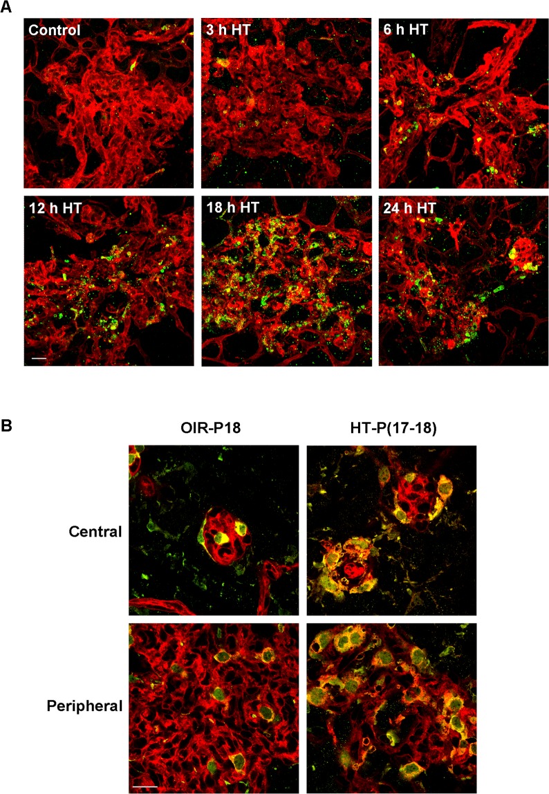
Figure 3. .
Hyperoxia treatment induces apoptosis in NV tufts and recruitment of macrophage/microglia. OIR mice were treated with hyperoxia (75% oxygen, HT) for 3 to 24 hours at P17. (A) Retinal flatmounts were stained with isolectin B4 (red) and anti-cleaved caspase-3 (green). Representative confocal images of NV tufts are shown (400×, n = 3 mice at each time point). Scale bar, 20 μm. (B) Retinal flatmounts were costained with isolectin B4 (red) for vessels and activated macrophage/microglia, and anti-Iba1 (green) for macrophage/microglia. Representative confocal images of NV tufts are shown (×630, n = 3 mice). Scale bar, 20 μm.

