Figure 2. .
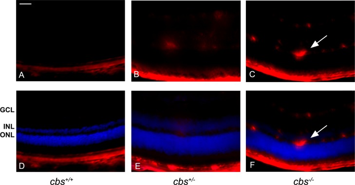
Increased endoglin (CD105) levels in cbs−/− mouse retinas. Fluorescent immunodetection of endoglin (red), a marker of neovascularization, was performed in retinal cryosections of 3-week-old mice; DAPI (blue) was used to label nuclei. Endoglin was minimally detected in cbs+/+ retinas (A, D), while endoglin levels were markedly increased in cbs−/− retinas (C, F), particularly in the ganglion cell layer and inner nuclear layer, where the blood vessels are predominant. Occasional endoglin-positive labeling was seen in cbs+/− retinas (B, E). The arrows represent neovascularization (new blood vessel formation). Scale bar: 50 μm. Experiments were performed in sections from six mice per group. Abbreviations for retinal layers are the same as for Figure 1.

