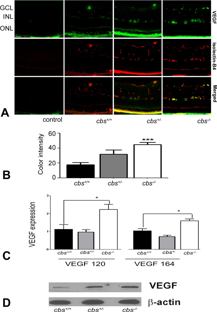
Figure 3. .
Increased VEGF levels in cbs−/− mouse retinas. (A) Fluorescent immunodetection of VEGF (green), a marker of neovascularization, and isolectin-B4 (red), a marker of endothelial cells, was performed in retinal cryosections from 3-week cbs+/+, cbs+/−, and cbs−/− mice. The far left panel provides controls: an isotype (IgM) control for VEGF and a negative control (omission of the primary antibody), but with inclusion of the secondary antibody, for isolectin-B4. Scale bar: 50 μm. Abbreviations for retinal layers are the same as for Figure 1. (B) Quantification of the intensity levels of VEGF immunofluorescence, showing significantly greater VEGF levels in cbs−/− compared to cbs+/+ and cbs+/− mice (***P < 0.001, n = 6). (C) Expression of VEGF mRNA is increased in neural retinas of cbs−/− mice compared to cbs+/− and cbs+/+ mice. Fold change in expression of vegf120 and vegf164 was determined by quantitative RT-PCR (*P < 0.05). (D) Representative immunoblot to detect VEGF in retinas of cbs+/+ mice compared with cbs+/− and cbs−/− mice. Protein was extracted from retina and subjected to SDS-PAGE; immunoblotting was performed with an affinity-purified antibody against VEGF 164 and subsequently with an antibody against β-actin (internal loading control).

