Figure 4. .
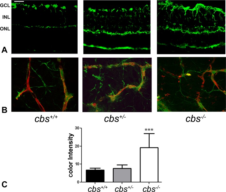
Increased GFAP expression in cbs−/− mouse retina. GFAP levels were detected by immunofluorescence in retinal cryosections and flat-mount preparations. (A) Retinal cryosections of 3-week cbs+/+, cbs+/−, and cbs−/− mice that were incubated with an antibody against GFAP followed by incubation with Alexa Fluor 488 (green)–labeled secondary antibody, showing increase in GFAP immune reactivity in both astrocytes and Müller cells in the cbs−/− retina compared to wild-type control cbs+/+, where GFAP is expressed in astrocytes only. (B) Retinal flat-mount preparations from cbs+/+, cbs+/−, and cbs−/− mice immunostained for isolectin-B4 (red) to visualize vasculature and GFAP (green), showing altered vasculature and ragged appearance of the astrocytes in cbs−/− mice compared to normal-shaped vasculature and astrocytes in the wild-type control cbs+/+ (n = 6). Scale bar: 50 μm. (C) Quantification of the data obtained from metamorphic analysis of color intensity of GFAP (significantly greater than wild-type, ***P < 0.001, n = 6).

