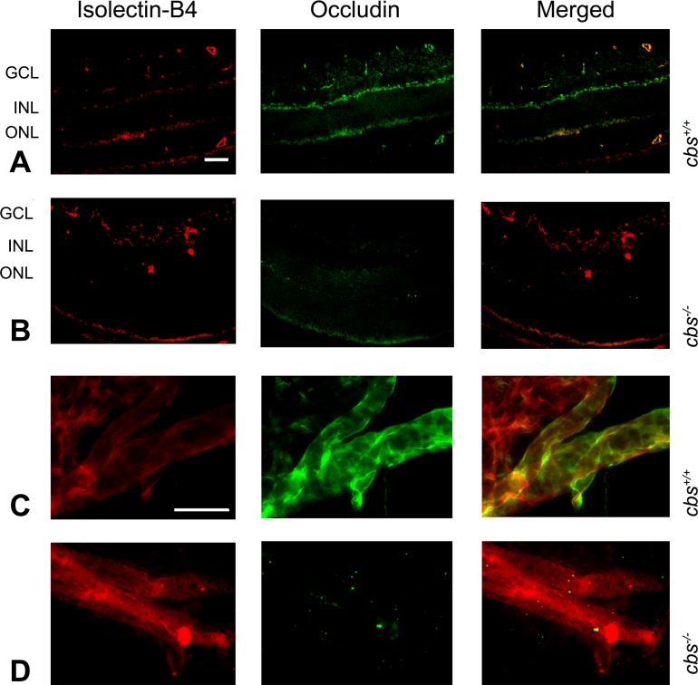
Figure 6. .
Occludin expression in mouse retina. Retinal cryosections of eyes from cbs+/+ and cbs−/− mice, incubated with antibodies against occludin and isolectin-B4 followed by incubation with Alexa Fluor 488 (green)– and Texas Red avidin (red)–labeled secondary antibodies, show decreased occludin immune reactivity in the cbs−/− retina (B) compared to wild-type cbs+/+ (A). Trypsin-digested retina, immunostained for isolectin-B4 (red) and occludin (green), show decreased occludin expression in cbs−/− retina (D) compared to cbs+/+ (C). Scale bar: 50 μm (A, B), 20 μm (C, D).

