
Figure 1.
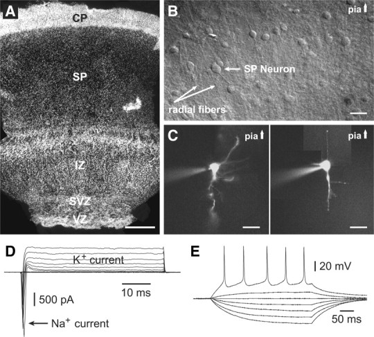
Human subplate zone at midgestation. A, Hoechst staining of the human fetal cerebral cortex at 20 gw–occipital lobe. Scale bar, 500 μm. VZ, Ventricular zone; SVZ, subventricular zone; IZ, intermediate zone. B, DIC image of live human cortical slice containing SP zone, SP neurons, and radial fibers (arrows). Scale bar, 20 μm. C, Rhodamine-filled SP neurons with long neurites pointing either toward SVZ (left) or toward the pial surface (right). Scale bar, 25 μm. D, Inward sodium and outward potassium voltage-gated currents in a human SP neuron. E, Repetitive action potential firing upon direct current injection.