
Figure 3.
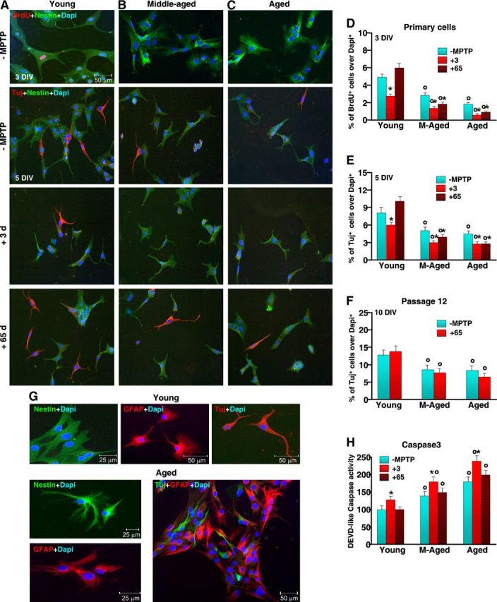
Aging primes SVZ cells to neurotoxin-induced long-lasting impairment of proliferative potential and neuroblast formation in vitro. NPCs were acutely isolated from the SVZ of 2–5-month-old (A), 8–10-month-old (B) and 22–24-month-old (C) mice, at both 3 and 65 d (7–8 mice/tp) after saline or MPTP injection, and cells processed as described. Proliferation (D) and neuron differentiation were studied in primary neurospheres (E) and after expansion for 12 passages, in vitro (F). A–C, Representative images comparing primary NPCs isolated from young (A), middle-aged (B), and aged (C) naive (−MPTP) mice and 3 and 65 dpt. Dual labeling with nestin (green) and BrdU (red) or nestin (green) and Tuj1 (red) counterstained with the nuclear marker, DAPI (blue) shows reduced BrdU+ and Tuj1+ cells with age and failure to recover upon exposure to MPTP (B, C), compared with young NPCs (A). D–F, NPC proliferation (D) as assessed by BrdU at 3 DIV; neuron differentiation, as assessed by Tuj1, after 5 DIV in primary cells (E); and neuron differentiation, as assessed by Tuj1, after 10 DIV, in expanded cells (F). G, Representative images of NPC isolated from young and aged mice after in vitro expansion. The cells were let to differentiate and stained with nestin (green), GFAP (red), Tuj1 (red), or with GFAP (red) and Tuj1 (green), counterstained with DAPI (blue). H, Comparison of cell death as assessed by Caspase3-like activity using the fluorogenic substrate DEVD-AFC as a function of age and MPTP. Results expressed as percentage changes relative to control (young −MPTP). *p < 0.05 versus −MPTP; °p < 0.05 versus young, within each respective group. The means ± SEM from three individual experiments is shown.