
Figure 5.
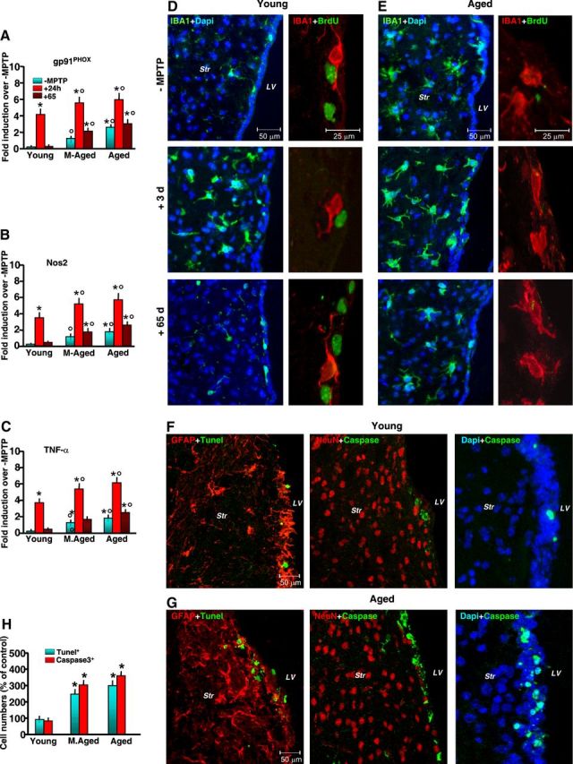
Aging exacerbates gpPhox, Nos2, and TNF-α mRNAs associated with microglial activation and cell-death marker expression in SVZ in response to MPTP. A–C, Comparison of quantified real-time PCR data using specific primers for gp91Phox, Nos2, and TNF-α in SVZ samples (6 mice/tp) from young, middle-aged, and aging mice in the absence or in the presence of MPTP treatment. The means ± SEM from three individual experiments are shown. Differences were analyzed by ANOVA followed by Newman–Keuls test, and considered significant when p < 0.05. *p < 0.05 versus saline; °p < 0.05 versus young within each experimental group. D, E, Representative confocal images showing dual staining with IBA1 (green) and DAPI (blue), and with IBA1 (red) and BrdU (green), in young (D) and aged (E) mice without (−MPTP) and 3 and 65 dpt. F–H, Dual localization of the death marker TUNEL (green) with GFAP (red), Caspase3 (green) with NeuN (red), and Caspase3 (green) with DAPI (blue) in young (F) and aged (G) mice show increased TUNEL+ and Caspase3+ cells and percentages of TUNEL+ and Caspase3+ cells (6 mice/tp, mean ± SD) in SVZ of aging compared with younger mice (H). *p < 0.05 versus young.