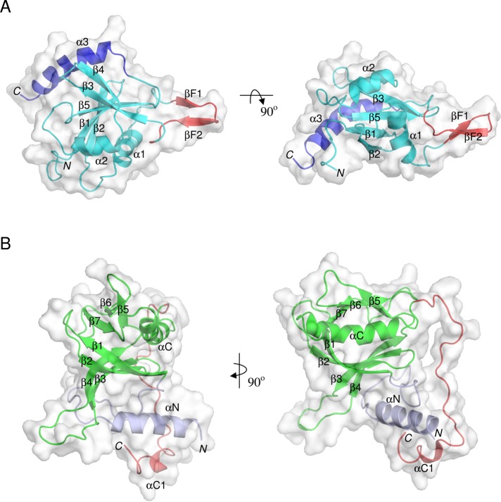
FIGURE 2:
Overall structures of the RBLSte11 and PHSte5 domains. (A) Solution NMR structure of the RBLSte11 domain. Canonical ubiquitin-like fold is in cyan, the inserted β-hairpin in red, and the helical C-terminal extension in blue. (B) Modeled PHSte5 domain. Canonical PH fold is in green and N- and C-terminal extensions are in light blue and red, respectively. Secondary structure elements are labeled. Two orthogonal views are presented in each case.

